Digital Cytogenetics at Krishna IVF: How IKAROS Supports Modern Chromosome Analysis
Looking Beyond Hormones, Eggs and Sperm
Infertility is usually caused by a combination of biological factors. Age, ovarian reserve, sperm production, reproductive anatomy, endocrine function and embryo development are all important. In some individuals and couples, however, an alteration in the number or structure of chromosomes may also contribute to infertility, recurrent pregnancy loss or the risk of a chromosomal condition in a pregnancy.
Cytogenetics is the branch of laboratory medicine that studies chromosomes. At Krishna IVF, cytogenetic evaluation is integrated with reproductive-medicine assessment, genetic counselling and digital chromosome-analysis technology.
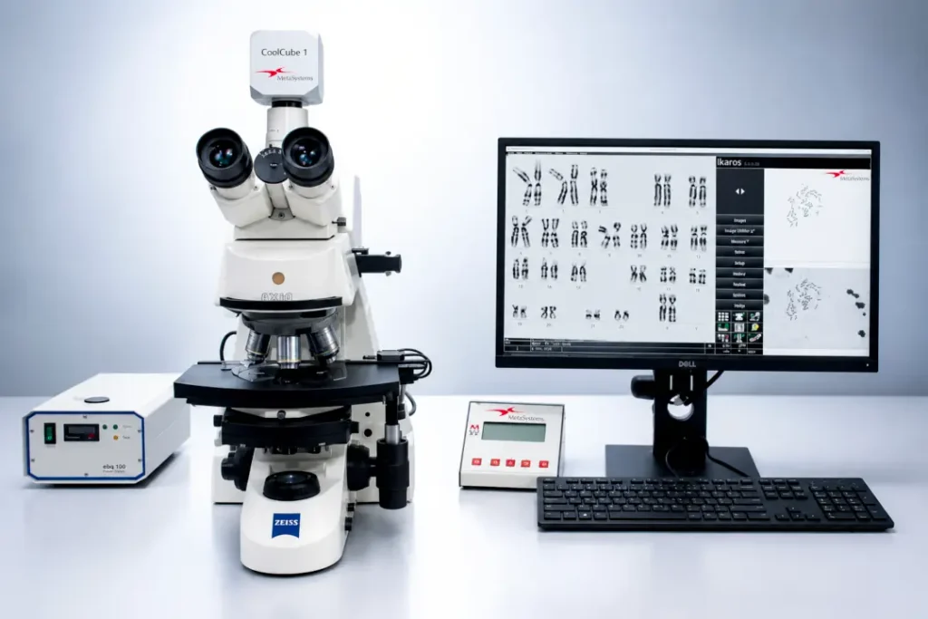
Krishna IVF uses IKAROS by MetaSystems to support digital karyotyping. The platform helps laboratory professionals process chromosome images, separate individual chromosomes and prepare a preliminary karyogram for expert review. The technology improves workflow efficiency, but it does not replace the judgement of a trained cytogenetics professional.
What Are Chromosomes?
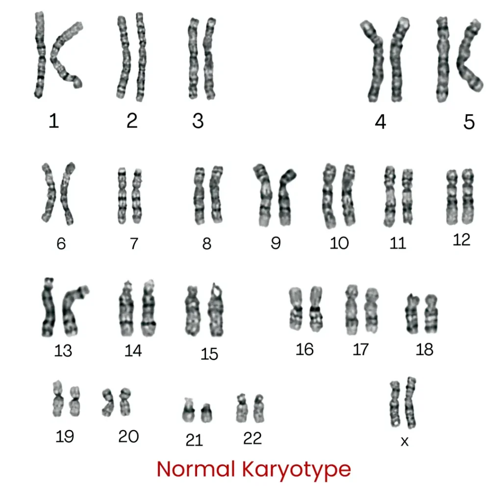
Chromosomes are organised structures made of DNA and proteins. Most human cells normally contain 46 chromosomes arranged into 23 pairs. One chromosome in each pair is usually inherited from each biological parent.
A change in chromosome number or structure may affect:
- Formation of eggs or sperm
- Fertilisation
- Embryo development
- Implantation
- Pregnancy continuation
- The health of a pregnancy or child
Some chromosome abnormalities cause a visible gain or loss of genetic material. Others are balanced rearrangements, in which chromosome material has changed position without an obvious net gain or loss.
A person with a balanced translocation or inversion may be completely healthy. However, some of that person’s eggs or sperm may contain an unbalanced amount of chromosome material. This may increase the likelihood of infertility, pregnancy loss or an affected pregnancy.
What Is a Karyotype?
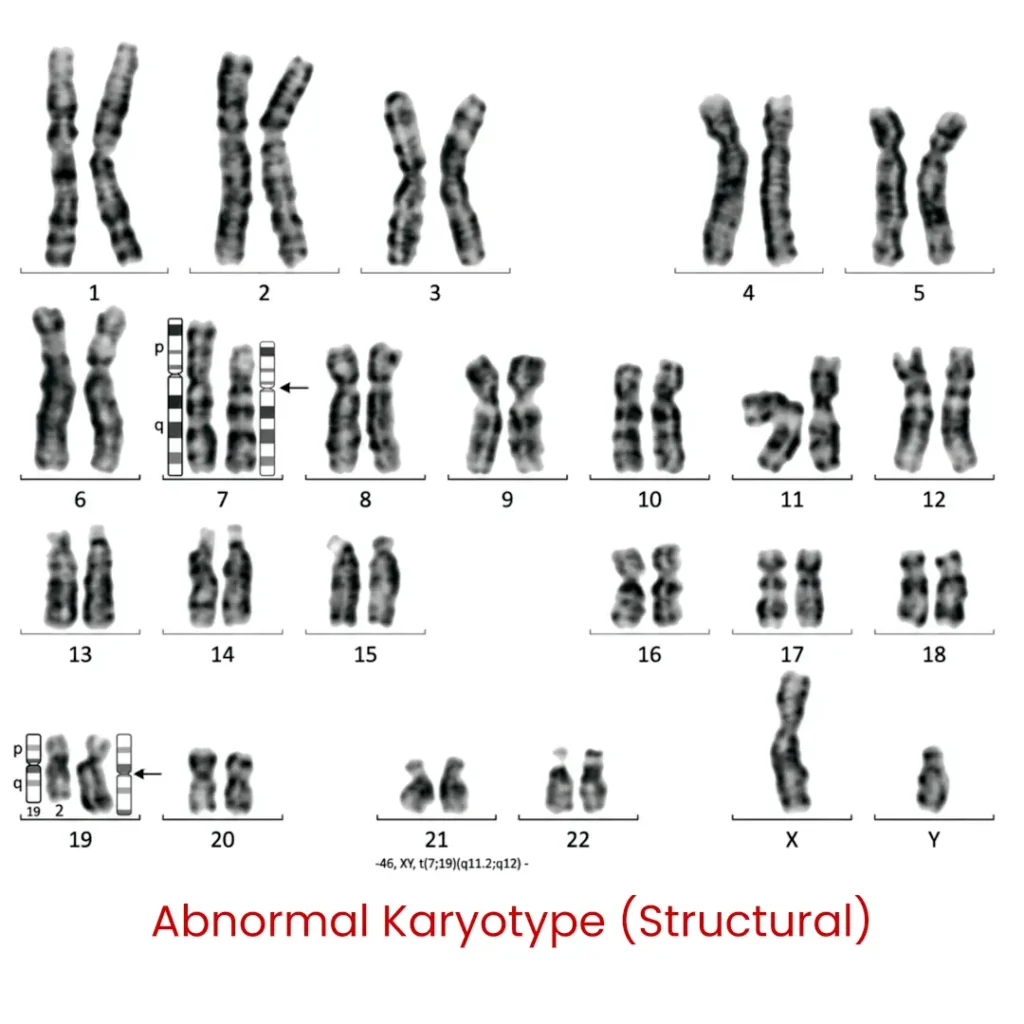
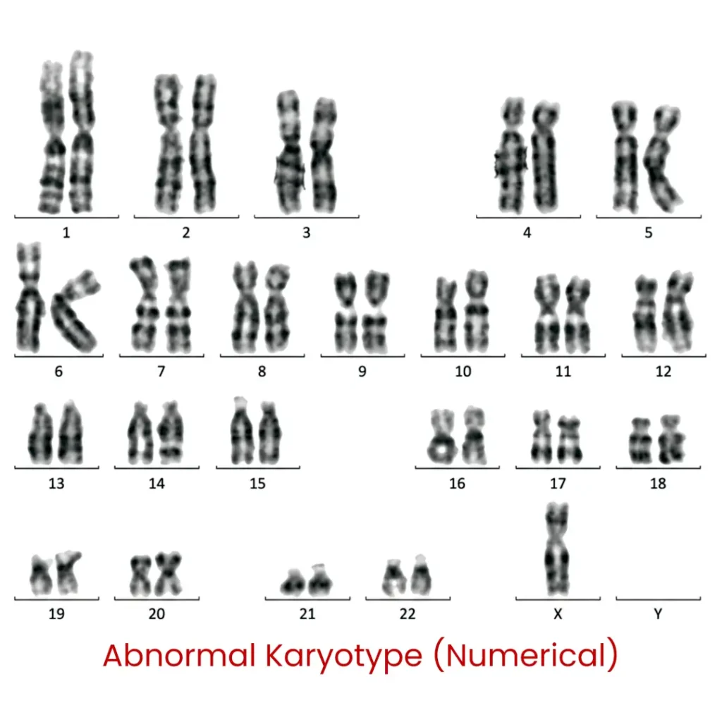
A karyotype is an organised representation of a person’s chromosomes. Chromosomes are examined under a microscope and arranged according to their size, banding pattern and centromere position.
Karyotyping can identify certain:
- Additional or missing chromosomes
- Balanced and unbalanced translocations
- Chromosomal inversions
- Large deletions or duplications
- Marker chromosomes
- Sex-chromosome abnormalities
- Forms of chromosomal mosaicism
Karyotyping provides a broad, genome-wide view of chromosomes. Its principal strength is the detection of numerical abnormalities and relatively large structural rearrangements, including balanced rearrangements that may not be identified by standard chromosomal microarray testing. Conventional karyotyping has lower resolution than molecular tests and cannot identify every small DNA alteration or single-gene disorder.
When May Karyotyping Be Recommended in Fertility Care?
Karyotyping is not required for every individual undergoing fertility evaluation or IVF. It is most useful when a person’s clinical history, reproductive history, physical findings or laboratory results suggest a higher probability of a chromosome abnormality.
1. Azoospermia and severe oligozoospermia
Chromosome abnormalities are more frequent among men with severely impaired sperm production than among men with normal sperm parameters.
The updated AUA–ASRM male-infertility guideline recommends karyotype testing for men with primary infertility and azoospermia or a sperm concentration below 5 million/mL when this is accompanied by elevated follicle-stimulating hormone, testicular atrophy or a clinical diagnosis of impaired sperm production.
Depending on the clinical findings, the evaluation may also include Y-chromosome microdeletion testing or other selected genetic investigations.
The results may help clinicians:
- Understand the possible cause of impaired sperm production
- Assess reproductive and genetic implications
- Counsel the couple before sperm retrieval or assisted reproduction
- Discuss the possibility of transmission to offspring
- Select appropriate additional investigations
2. Recurrent pregnancy loss
Many first-trimester pregnancy losses are caused by sporadic chromosome abnormalities in the embryo. In a smaller proportion of couples, one partner may carry a balanced structural chromosome rearrangement.
Current recommendations support a targeted approach to parental karyotyping. Testing is especially relevant when pregnancy-tissue analysis identifies an unbalanced structural chromosome abnormality or when pregnancy tissue was unavailable for genetic testing. Individual reproductive history and family history should also inform the decision.
Identifying a parental rearrangement can help guide genetic counselling and discussion of future reproductive options. It does not, by itself, determine whether a couple can achieve a healthy pregnancy.
3. Primary ovarian insufficiency or amenorrhoea
Selected individuals with primary ovarian insufficiency, absent or delayed puberty, primary amenorrhoea or clinical features suggesting gonadal dysgenesis may require chromosome analysis as part of a wider diagnostic evaluation.
The decision should be based on age, menstrual history, endocrine results, reproductive anatomy and associated clinical findings. Cytogenetic testing in this context is intended to support medical diagnosis and reproductive counselling. It must never be used for non-medical sex determination or sex selection.
4. A known chromosome rearrangement in the family
Karyotyping may be considered when:
- A parent, sibling or other close relative carries a translocation or inversion
- A previous child or pregnancy had a structural chromosome abnormality
- Pregnancy-tissue testing demonstrated an unbalanced rearrangement
- A couple is considering preimplantation genetic testing for structural rearrangements, or PGT-SR
5. Selected disorders of reproductive development
Chromosome analysis may contribute to the multidisciplinary evaluation of individuals with differences in pubertal development, gonadal function or reproductive anatomy when a chromosome abnormality is clinically suspected.
How Is a Karyotype Prepared?
Karyotyping is a multistep laboratory process. The final karyogram represents only one part of the complete workflow.
Step 1: Clinical assessment
The fertility specialist or clinical geneticist first determines whether karyotyping is appropriate. The patient should receive an explanation of:
- Why the test is being requested
- What it may identify
- What it cannot identify
- How the result may affect the individual or biological relatives
- Whether genetic counselling may be required
Step 2: Sample collection
Peripheral blood is commonly used for constitutional karyotyping. Correct patient identification, sample labelling, transport conditions and traceability are essential.
Step 3: Cell culture
Lymphocytes from the blood sample are cultured under controlled laboratory conditions. The purpose is to obtain actively dividing cells.
Step 4: Metaphase preparation
Cell division is arrested during metaphase, when chromosomes are condensed and can be examined microscopically. The cells are harvested, placed on slides and stained to create characteristic chromosome banding patterns.
Step 5: Digital image acquisition
Suitable metaphase cells are selected, and chromosome images are acquired through the laboratory’s validated microscope and digital imaging workflow.
Image quality depends on several factors, including:
- Chromosome spreading
- Banding quality
- Chromosome contraction
- Overlapping or touching chromosomes
- Background artefacts
- Focus and image contrast
Step 6: Karyogram preparation using IKAROS
The metaphase image is transferred into the IKAROS environment. The software supports image processing, chromosome segmentation, chromosome classification and preparation of a preliminary karyogram.
MetaSystems describes newer IKAROS workflows as using deep neural networks to support chromosome separation, classification, overlap estimation and band-resolution assessment. Software-generated karyogram proposals remain drafts until they are reviewed and explicitly approved by the user.
The exact functions available at Krishna IVF depend on the installed software version, licensed modules, microscope configuration and validated local workflow.
Step 7: Expert analysis
A trained cytogenetics professional evaluates:
- Chromosome number
- Chromosome morphology
- Banding patterns
- Possible structural rearrangements
- Consistency across analysed cells
- Evidence of mosaicism or more than one cell line
Additional metaphases may be examined when an abnormality is suspected or when the initial cells are technically inadequate.
Step 8: Reporting and clinical interpretation
The result is documented using accepted cytogenetic nomenclature and interpreted in the context of the patient’s phenotype, reproductive history and other investigations.
A chromosome result should not be interpreted in isolation. Genetic counselling may be required before reproductive decisions are made.
How IKAROS Supports the Cytogenetics Laboratory
Traditional karyotyping involved photographing metaphase cells, physically cutting out chromosome images and arranging them manually. Digital systems have transformed this process.
IKAROS provides tools for:
- Digital chromosome-image acquisition and processing
- Image enhancement and contrast adjustment
- Separation of individual chromosomes
- Preliminary chromosome classification
- Arrangement into a draft karyogram
- Band-resolution assessment in supported versions
- Case documentation and image archiving
- Review and correction before authorisation
MetaSystems states that IKAROS is designed to reduce the number of manual interactions required during karyogram preparation and may decrease the time needed for analysis and review compared with entirely manual karyotyping.
Potential laboratory benefits
Greater workflow efficiency
Repetitive image-processing and chromosome-arrangement tasks can be completed more quickly, allowing laboratory professionals to concentrate on abnormality recognition and interpretation.
Improved standardisation
Digital tools support a more consistent approach to image display, chromosome arrangement and case documentation.
Easier review
Chromosome images and prepared karyograms can be reviewed, corrected and discussed without physically reconstructing the case.
Better traceability
Digital records can support internal quality review, teaching, audit and authorised second opinions, subject to institutional privacy and access-control policies.
Why Human Review Cannot Be Replaced
Chromosomes in a metaphase spread are rarely presented as perfectly separated, straight structures. They may:
- Touch or overlap
- Bend or twist
- Break during preparation
- Appear highly contracted
- Have weak or uneven banding
- Resemble another chromosome
- Contain subtle structural abnormalities
An algorithm may separate one chromosome into two fragments, combine two chromosomes, assign a chromosome to an incorrect pair or fail to recognise the clinical importance of a subtle banding change.
For this reason, an automatically prepared karyogram is a draft, not a final diagnosis. MetaSystems specifies that automated suggestions become part of official case information only after user approval.
At Krishna IVF, IKAROS functions as a laboratory-support platform. Final interpretation and report authorisation remain professional responsibilities.
Karyotype, Microarray, FISH and PGT: Different Tests for Different Questions
| Investigation | What it principally evaluates | Important strength | Important limitation |
|---|---|---|---|
| Conventional karyotype | Number and visible structure of chromosomes | Detects many balanced rearrangements and provides cell-level analysis | Cannot detect many small DNA changes |
| Chromosomal microarray | Submicroscopic gains and losses of DNA | Higher resolution for copy-number changes | Usually cannot detect balanced translocations or inversions |
| FISH | Selected chromosome regions | Targeted and relatively rapid | Only examines regions covered by the selected probes |
| PGT-SR | Embryos from carriers of structural chromosome rearrangements | Helps identify embryos without an unbalanced rearrangement detectable by the assay | Requires IVF and does not guarantee an embryo suitable for transfer or a live birth |
| PGT-A | Chromosome copy-number findings in embryo-biopsy samples | May provide embryo chromosome information in selected clinical situations | Does not diagnose every genetic condition or guarantee implantation and live birth |
No single technology identifies every type of genetic abnormality. Test selection should begin with the clinical question rather than with the perceived sophistication of the platform.
What Happens if a Chromosome Abnormality Is Found?
An abnormal karyotype does not necessarily mean that pregnancy is impossible.
The next steps depend on the exact chromosome finding and may include:
- Confirmation and detailed interpretation
- Karyotyping of the reproductive partner
- Testing of selected relatives
- Formal genetic counselling
- Discussion of natural conception and prenatal diagnostic options
- IVF with PGT-SR in selected couples
- Consideration of donor gametes where appropriate
- Psychological support and time for informed decision-making
For balanced translocation carriers, reproductive outcomes vary according to the chromosomes involved, the breakpoint positions, whether the carrier is male or female, maternal age, ovarian reserve and the number of embryos available. Evidence comparing PGT-SR with expectant management remains limited by heterogeneity and the absence of strong prospective trials.
Ethical, Private and Patient-Centred Cytogenetics
Chromosome results can have implications beyond the person tested. A finding may provide information about parents, siblings, children or other biological relatives.
Cytogenetic services must therefore maintain:
- Confidential sample handling
- Secure storage of digital chromosome images
- Controlled access to genetic information
- Documented reporting and authorisation
- Appropriate pre-test and post-test counselling
- Sensitive communication of uncertain or unexpected findings
- Compliance with applicable Indian laws and professional standards
Chromosome analysis in reproductive medicine must be used for legitimate medical indications. It must not be presented or used as a pathway for non-medical sex selection or fetal sex determination.
The Krishna IVF Approach
Krishna IVF integrates cytogenetics with reproductive medicine, andrology, embryology and reproductive genetics.The objective is not simply to produce a chromosome image. It is to connect the laboratory result with the clinical question:
- Why is the patient experiencing impaired sperm production?
- Could a chromosome rearrangement contribute to pregnancy loss?
- Does the finding affect the safety or interpretation of fertility treatment?
- Is further molecular testing required?
- Would genetic counselling or PGT-SR be clinically relevant?
- What reproductive options should be discussed with the couple?
IKAROS supports this process by improving digital chromosome handling and karyogram preparation. It does not promise pregnancy, prevent every pregnancy loss or guarantee that an embryo will be chromosomally normal.
Conclusion
Cytogenetic evaluation can provide clinically important information for selected individuals and couples with severe male-factor infertility, recurrent pregnancy loss, suspected sex-chromosome abnormalities or a family history of chromosome rearrangements.
At Krishna IVF, IKAROS by MetaSystems supports digital chromosome-image processing, chromosome segmentation and draft karyogram preparation. Each result must still undergo expert review, correction where required and professional authorisation.
The responsible model for modern cytogenetics is therefore not automation in place of expertise. It is:
Digital technology supporting careful laboratory practice, clinical interpretation and informed reproductive decision-making.
Frequently Asked Questions
Is karyotyping necessary before every IVF cycle?
No. Karyotyping is recommended only when there is an appropriate clinical or genetic indication.
Can a normal karyotype exclude every genetic condition?
No. A normal karyotype does not exclude small deletions, small duplications, single-gene disorders or many DNA-sequence variants.
Does an abnormal karyotype mean that a person cannot have a healthy child?
No. Reproductive possibilities depend on the exact chromosome finding, age, ovarian reserve, sperm production and previous reproductive history. Individual genetic counselling is essential.
Does IKAROS make the final diagnosis?
No. IKAROS supports digital image processing and draft karyogram preparation. The final analysis and report must be reviewed and authorised by trained professionals.
Is a karyotype the same as PGT?
No. A karyotype generally analyses chromosomes from an individual’s cells. PGT analyses a small sample of cells biopsied from an embryo created through IVF.
Can karyotyping guarantee a successful IVF outcome?
No. Karyotyping provides genetic information. IVF outcomes continue to depend on many clinical, biological and embryological factors.
Medical Disclaimer
This article is intended for general educational purposes. It is not a substitute for individual medical evaluation, genetic counselling, diagnosis or treatment planning. The appropriate genetic test depends on the patient’s clinical history, reproductive findings and family history. Fertility-treatment outcomes vary among individuals.
References
- MetaSystems. Ikaros karyotyping software. MetaSystems International; 2026. Accessed August 6, 2026.
- MetaSystems. Quick guide: Ikaros 6.3. MetaSystems International; 2024.
- American Urological Association; American Society for Reproductive Medicine. Diagnosis and treatment of infertility in men: AUA/ASRM guideline. Amended 2024.
- American Society for Reproductive Medicine Practice Committee. Recurrent pregnancy loss: a committee opinion. Fertil Steril. 2026;125:1023-1041.
- ESHRE Guideline Group on Recurrent Pregnancy Loss. ESHRE guideline: recurrent pregnancy loss—update 2022. Hum Reprod Open. 2023;2023(1):hoad002.