Fluorescence in situ hybridization (FISH) is a molecular cytogenetic laboratory technique used to identify selected chromosomal abnormalities using fluorescently labeled DNA probes.
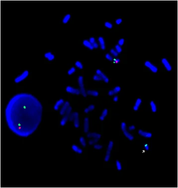
In this technique, specially designed DNA probes bind to complementary chromosomal regions within interphase nuclei or metaphase chromosomes. The fluorescent signals are then visualized using a fluorescence microscope equipped with appropriate optical filters.
FISH analysis may assist identification of selected numerical chromosomal abnormalities (aneuploidies), structural rearrangements, microdeletions, translocations, and other chromosomal variations depending on the clinical indication and probe selection.
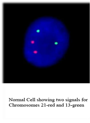
In reproductive and prenatal evaluation workflows, FISH may be utilized in selected situations for assessment of chromosomes commonly associated with aneuploidy including chromosomes 13, 18, 21, X, and Y.
FISH techniques may additionally be utilized in selected hematological, oncological, prenatal, cytogenetic, and post-transplant evaluation workflows depending on the clinical condition and laboratory requirements.
Different sample types may be utilized for FISH analysis including peripheral blood, amniotic fluid, bone marrow, tissue samples, or other cytogenetic specimens depending on the indication for testing.
The laboratory workflow generally involves specimen preparation, probe hybridization, fluorescence signal detection, microscopic evaluation, and qualitative or quantitative interpretation of fluorescent signal patterns.
FISH testing at Krishna IVF forms part of broader reproductive genetics, molecular cytogenetics, prenatal evaluation, laboratory diagnostics, and individualized reproductive medicine workflows.