Ultrasound-Guided Embryo Transfer
Embryo transfer is a procedure performed as part of assisted reproductive treatment in which an embryo or embryos are placed into the uterine cavity using a specialized transfer catheter. The procedure is coordinated between the clinician and the embryology team according to the individual treatment plan.
Role of Ultrasound Guidance
Ultrasound imaging enables the clinician to view the uterus and follow the position of the transfer catheter during the procedure. It may assist in assessing:
- Uterine position and orientation
- Cervical anatomy and catheter pathway
- Position of the catheter tip
- Intended area of embryo deposition
- Distance from the uterine fundus
These findings are considered together with the patient’s clinical history, previous imaging and treatment requirements.
Transabdominal and Transvaginal Guidance
Ultrasound guidance during embryo transfer may be performed using either a transabdominal or transvaginal approach.
During transabdominal ultrasound guidance, the probe is placed over the lower abdomen. A suitably filled bladder may be required to improve visualization.
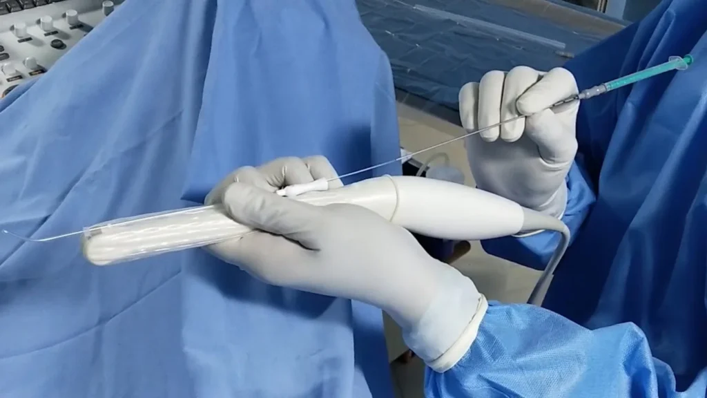
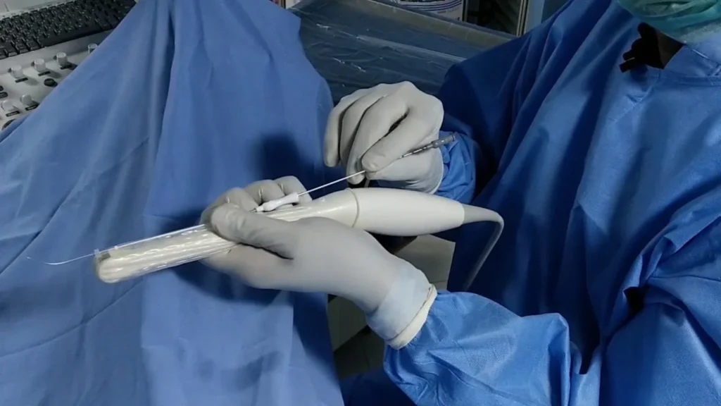
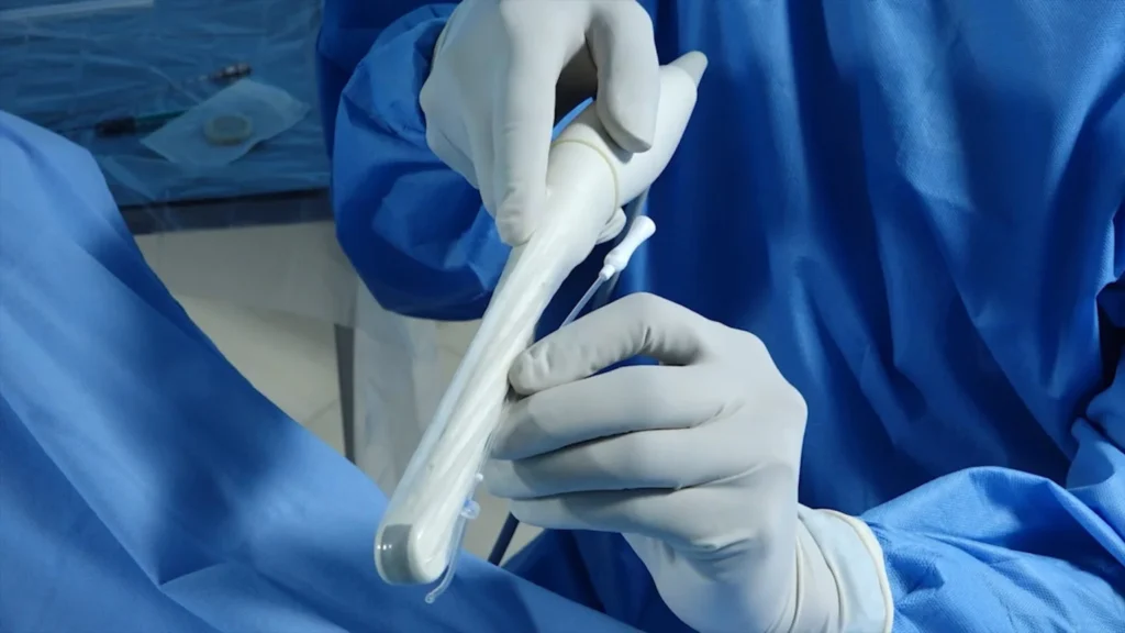
Transvaginal ultrasound guidance uses a covered vaginal ultrasound probe to provide imaging closer to the uterus. It generally does not require a full bladder and may provide clearer visualization in certain circumstances, including some uterine positions or when transabdominal imaging is limited.
The appropriate approach is selected according to uterine and cervical anatomy, previous transfer experience, procedural requirements and clinical assessment.
Transvaginal Ultrasound-Guided Transfer
Krishna IVF Clinic performs transvaginal ultrasound-guided embryo transfer where considered clinically appropriate.
During the procedure, an outer catheter is carefully positioned through the cervix. A mock transfer may be performed to assess the catheter pathway before the embryo-transfer catheter is introduced.
The embryologist loads the selected embryo or embryos into the transfer catheter. Using real-time ultrasound imaging, the clinician follows the catheter tip and deposits the embryo or embryos at the planned position within the uterine cavity. The catheter is then returned to the embryologist to confirm that the transfer is complete.
Ultrasound images or video recordings may be retained as part of procedural documentation and review.
Patient Information
Transvaginal ultrasound guidance avoids the need to maintain a full bladder. Embryo transfer is generally a brief procedure, although its duration and the level of discomfort may vary depending on individual anatomy and procedural circumstances.
Anaesthesia is not routinely required but may be considered in selected situations based on clinical requirements.
Ultrasound guidance supports visualization and catheter placement but cannot guarantee implantation, pregnancy or a particular treatment outcome. Outcomes depend on several factors, including embryo characteristics, age, reproductive and uterine factors and the overall treatment protocol.