
How Artificial Intelligence Is Changing Follicle Monitoring in IVF
From our early 3D ultrasound research to a new international study
Krishna IVF, Visakhapatnam | September 2026
When a woman undergoes IVF treatment, one of the most familiar parts of the process is the ultrasound scan used to monitor growing follicles in the ovaries.
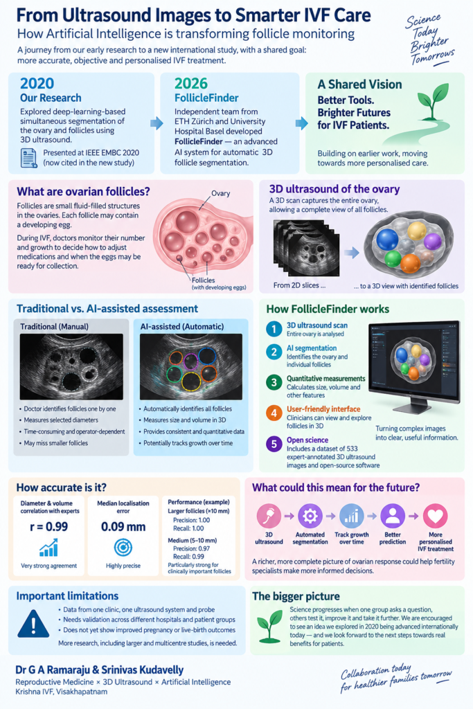
Doctors measure these small fluid-filled structures because each follicle may contain a developing egg. Their number and growth help the fertility team decide how to adjust medications and when the eggs may be ready for collection.
Traditionally, this requires the doctor to identify and measure follicles individually.
But could a computer analyse the whole ovary in three dimensions, identify every follicle and measure them automatically?
That was a question our collaborative research began exploring several years ago.
Our work in 2020
In 2020, our team investigated whether artificial intelligence (AI) and deep learning could analyse 3D transvaginal ultrasound images and automatically identify both the ovary and the follicles within it.
Our work was presented at the IEEE Engineering in Medicine and Biology Society conference and has now been cited in a new international study.
s41598-026-69790-y_reference.pdf
For us, this new citation is meaningful because it shows that the scientific question we were exploring then continues to be developed by independent researchers today.
Six years later: FollicleFinder
Researchers from ETH Zürich, the Swiss Institute of Bioinformatics and University Hospital Basel have now reported a system called FollicleFinder in Scientific Reports.
Their goal is straightforward: use AI to identify and measure ovarian follicles automatically from 3D ultrasound images.
The researchers developed software that can analyse a 3D scan, separate individual follicles and calculate measurements such as their size and volume.
The researchers developed software that can analyse a 3D scan, separate individual follicles and calculate measurements such as their size and volume.
They have also created a user-friendly interface so that researchers and clinicians can view the follicles in three dimensions.
Importantly, they are making available a dataset containing 533 expert-annotated 3D ultrasound images, together with their open-source software.
Why could this matter for IVF patients?
During IVF stimulation, doctors repeatedly assess how a group of follicles is developing.At present, much of this assessment depends on measuring selected follicle diameters.
AI-assisted 3D ultrasound could eventually allow us to look at the entire group of growing follicles, rather than only a few measurements.
In the future, this could create a much richer picture of ovarian response:
3D ultrasound → automatic follicle measurement → tracking growth over time → better prediction → more personalised IVF treatment
The ultimate goal is not to replace the fertility specialist.
It is to give clinicians more consistent and quantitative information on which to base decisions.
How accurate is the new system?
The results are encouraging.
In the study, automated measurements of follicle diameter and volume were very strongly correlated with expert-defined measurements, with a reported correlation of r = 0.99. The median error in locating a follicle was only 0.09 mm.
The system performed particularly well in identifying larger follicles, which are important during treatment monitoring.
But these are measures of technical accuracy, not proof that using the system will increase pregnancy or live-birth rates.
That distinction is important.
What makes this personally meaningful to us?
Science rarely progresses because of one paper or one research group.
A team asks a question. Another group develops a better method. Larger datasets become available. Technology improves. Eventually, some of those ideas may find their way into everyday patient care.
The new paper specifically discusses our 2020 study while describing the development of deep-learning approaches for simultaneous 3D segmentation of the ovary and follicles.
Seeing an independent team from Switzerland take this scientific problem further six years later is therefore particularly satisfying.
It reminds us why collaboration between doctors and engineers is so valuable.
Doctors understand the clinical problem.
Engineers may see completely different ways of measuring, analysing and solving it.
When those two perspectives come together, important innovations can emerge.
Are we ready to use AI to make IVF decisions?
Not yet.The authors themselves identify important limitations. Their main images came from one clinic using one ultrasound machine and probe. The technology still needs validation across different hospitals, ultrasound systems and patient populations. Most importantly, this study did not demonstrate that AI-guided follicle measurement improves pregnancy or live-birth outcomes.
That should be the next stage of research.
For patients, therefore, the message is not that “AI can now manage IVF.”
The more accurate message is: AI may eventually help fertility specialists measure and understand ovarian response more consistently—but careful clinical validation must come first.
For Srinivas and me, it is rewarding to see a research question we explored in 2020 continuing to evolve.
And perhaps that is one of the most satisfying parts of scientific work: an idea does not have to remain yours to remain meaningful. Its real value appears when others test it, improve it and take it further.
Dr G A Ramaraju & Srinivas Kudavelly
Reproductive Medicine, 3D Ultrasound and Artificial Intelligence
References
- Franz L, Yamauchi KA, Fricke S, et al. FollicleFinder enables automated three-dimensional segmentation of human ovarian follicles. Scientific Reports. 2026. doi:10.1038/s41598-026-69790-y.
- Mathur P, Kakwani K, Kudavelly S, Ramaraju GA, et al. Deep learning based quantification of ovary and follicles using 3D transvaginal ultrasound in assisted reproduction. 42nd Annual International Conference of the IEEE Engineering in Medicine & Biology Society (EMBC). 2020:2109-2112.