
Standard G-Banded Karyotyping: Uses in Fertility and Pregnancy
Standard G-Banded Karyotyping: What This Chromosome Test Can Reveal About Fertility and Pregnancy
Chromosomes are organised packages of DNA found in nearly every cell of the body. Most people have 46 chromosomes arranged in 23 pairs. Changes in chromosome number or structure may be associated with infertility, recurrent pregnancy loss, congenital conditions or differences in reproductive development.
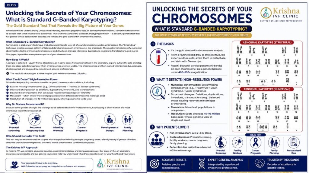
Standard G-banded karyotyping allows specialists to examine the number and broad structure of all chromosomes under a microscope. Newer genetic technologies can detect smaller DNA changes, but karyotyping remains particularly useful when the clinical question concerns whole-chromosome architecture, including balanced translocations and inversions.
What is standard G-banded karyotyping?
A karyotype is an organised picture of a personʼs chromosomes. The chromosomes are arranged in pairs according to their size, banding pattern and the position of the centromere—the narrowed region of each chromosome.
The term G-banded refers to the characteristic light and dark bands produced after the chromosomes are treated and stained with Giemsa. These bands act like a biological barcode. By comparing corresponding chromosomes, a trained cytogenetic scientist can identify important numerical or structural differences.
A typical result may be written as:
- 46,XX
- 46,XY
An abnormal result uses internationally standardised cytogenetic terminology to describe the chromosome involved and the nature of the change. The written result should not be interpreted as an isolated laboratory code. It must be considered alongside the personʼs medical history, fertility findings, previous pregnancies and family history.
How is the test performed?
For most adults undergoing constitutional chromosome testing, the sample is peripheral blood. Other samples may be used for specific clinical indications, including:
- Amniotic fluid
- Chorionic villi
- Pregnancy-loss tissue
- Bone marrow
- Skin or other tissues
In the laboratory, cells are cultured so that they begin to divide. Their division is then arrested during metaphase, the stage at which chromosomes are condensed and most clearly visible.
The cells undergo further processing so that the chromosomes spread across a microscope slide. After controlled enzyme treatment and staining with Giemsa, the characteristic G-bands become visible.
Digital images of suitable metaphase cells are captured. The chromosomes are then:
- Counted
- Arranged into corresponding pairs
- Compared according to their banding patterns
- Examined for numerical and structural abnormalities
Several cells are usually assessed. Additional cells may be studied when mosaicism is suspected.
Because living cells must first grow in culture, karyotyping generally takes longer than rapid molecular assays. Turnaround time varies according to the specimen, clinical indication, cell growth and laboratory workflow.
What does “band resolution” mean?
A karyotype report may mention a 400-, 550- or higher-band level. This describes how many bands can be distinguished across one complete haploid chromosome set.
It does not mean that the chromosomes were examined only at 400-times or 550-times microscope magnification.
Higher band levels generally allow more precise localisation of a structural chromosome change. However, even a technically good karyotype cannot identify every genetic abnormality.
Its practical resolution depends on:
- Specimen quality
- The chromosome region involved
- The size of the abnormality
- The type of structural change
- The quality of metaphase preparation
Small copy-number changes may fall below the resolution of conventional karyotyping and may require chromosomal microarray, targeted molecular testing or DNA sequencing.
What can G-banded karyotyping detect?
1. Numerical chromosome abnormalities
Karyotyping can identify an additional or missing chromosome. Examples include:
- Trisomy 21
- Monosomy X
- 47,XXY
- Other autosomal or sex-chromosome abnormalities
It may also detect larger numerical abnormalities such as triploidy, in which three complete chromosome sets are present instead of two.
2. Structural chromosome abnormalities
G-banded karyotyping may detect sufficiently large:
- Deletions
- Duplications
- Reciprocal translocations
- Robertsonian translocations
- Inversions
- Ring chromosomes
- Marker chromosomes
A balanced translocation occurs when chromosome segments exchange positions without an obvious net loss or gain of genetic material. The carrier may be healthy and may remain unaware of the rearrangement until infertility, recurrent miscarriage or an affected pregnancy occurs.
During the formation of eggs or sperm, however, a balanced rearrangement can produce cells with an unbalanced chromosome complement. This may contribute to:
- Failure of embryo development
- Pregnancy loss
- Reduced reproductive potential
- A pregnancy affected by a chromosome imbalance
The reproductive implications depend on the chromosomes involved and the exact positions of the breakpoints.
3. Mosaicism
Mosaicism means that two or more genetically different cell lines are present in the same person. For example, some cells may have a typical chromosome complement while others contain an abnormality.
The ability to detect mosaicism depends on:
- The proportion of affected cells
- The number of cells analysed
- The tissue tested
- Cell-culture behaviour
- The nature of the abnormality
A normal blood karyotype cannot completely exclude very low-level mosaicism or an abnormal cell line confined to another tissue.
Why may karyotyping be advised during fertility evaluation?
Karyotyping is not required for every person with infertility. It is most informative when the clinical picture indicates an increased probability of a chromosome abnormality.
Karyotyping in male infertility
Current AUA–ASRM guidance recommends karyotype testing for men with primary infertility and:
- Azoospermia; or
- A sperm concentration below 5 million/mL
This is particularly relevant when the findings are accompanied by elevated FSH, testicular atrophy or presumed impairment of sperm production.
A karyotype may identify a sex-chromosome abnormality or a balanced structural rearrangement. The result may influence counselling about:
- The cause of impaired sperm production
- The possibility of sperm retrieval
- The potential use of ICSI
- The possibility of transmitting the chromosome change
- The need for additional genetic testing
- Reproductive and prenatal testing options
Y-chromosome microdeletion analysis is a separate molecular test. It examines selected regions of the Y chromosome and does not replace karyotyping. Similarly, a normal karyotype does not exclude Y-chromosome microdeletions or pathogenic
variants in individual genes.
Karyotyping in recurrent pregnancy loss
Chromosome abnormalities are an important biological cause of pregnancy loss. The 2026 ASRM committee opinion recommends a stepwise evaluation that begins, when feasible, with chromosome assessment of miscarriage tissue, followed by further investigations according to the result and clinical history.
Parental karyotyping is not automatically required for every couple who has experienced pregnancy loss. ESHRE recommends considering parental chromosome testing after an individual risk assessment—for example, when:
- Pregnancy tissue shows a structural rearrangement
- There is a family history of a chromosome abnormality
- A previous pregnancy or child had congenital abnormalities
- Other clinical findings suggest a parental rearrangement
The likelihood of finding an abnormal parental karyotype is relatively low, but the information can be important when a clinically significant rearrangement is identified.
When either partner carries a balanced rearrangement, genetic counselling is essential. A healthy pregnancy may still occur naturally, but the probability of an unbalanced conception varies according to the particular rearrangement.
Other selected reproductive indications
Karyotyping may also be considered in selected patients with:
- Primary amenorrhoea
- Premature ovarian insufficiency
- Atypical pubertal development
- Differences of sex development
- Multiple congenital abnormalities
- A known familial chromosome rearrangement
The decision should follow an individual clinical assessment rather than being included automatically in a broad “genetic package.”
Karyotype, microarray, FISH and sequencing: how are they different?
No single genetic test can identify every type of abnormality.
| Test | Main strength | Important limitation |
|---|---|---|
| G-banded karyotype | Examines chromosome number, large structural changes and many balanced rearrangements | May miss small or submicroscopic changes |
| Chromosomal microarray | Detects smaller gains and losses of DNA | Usually cannot identify truly balanced rearrangements |
| FISH | Rapid, targeted examination of selected chromosome regions | Only assesses the regions targeted by the probes |
| Gene panel or sequencing | Detects sequence-level variants in selected genes or across the genome | Does not replace cytogenetic testing for every structural chromosome abnormality |
Chromosomal microarray provides greater resolution for detecting small deletions and duplications. However, it may not reveal a balanced translocation or show the physical architecture of the chromosomes in the same way as a karyotype.
FISH uses fluorescent probes to examine particular chromosome regions. It is useful when a specific abnormality is suspected or rapid targeted information is required, but it is not a complete genome-wide assessment.
Gene panels, exome sequencing and genome sequencing examine DNA at the sequence level. They may identify pathogenic variants in individual genes but do not automatically replace conventional cytogenetic analysis.
In prenatal diagnosis, chromosomal microarray is particularly useful when ultrasound identifies one or more major fetal structural abnormalities. When the fetus is structurally normal and invasive diagnostic testing is undertaken, karyotyping or microarray may be selected according to the clinical question and pre-test counselling.
What does a normal karyotype not exclude?
A normal karyotype is reassuring regarding visible chromosome number and structure, but it does not rule out:
- Small deletions or duplications below the testʼs resolution
- Single-gene disorders
- Most DNA sequence variants
- Epigenetic conditions
- Low-level mosaicism
- Abnormalities confined to an untested tissue
- Every genetic cause of infertility or miscarriage
A normal report therefore does not mean that “all genetic tests are normal.” It means that no abnormality detectable by that particular method was identified.
What happens if the result is abnormal?
An abnormal result should be reviewed with a reproductive-medicine specialist and a clinical geneticist or genetic counsellor.
The discussion may include:
- Whether the change is balanced or unbalanced
- Whether the finding explains the clinical problem
- Whether the partner or other relatives require testing
- The estimated reproductive implications
- Options for future pregnancies
- The limitations of available genetic tests
Depending on the diagnosis, reproductive pathways may include natural conception with prenatal diagnosis, IVF with preimplantation genetic testing for structural rearrangements—PGT-SR, donor gametes or other family-building options.
PGT-SR may help identify embryos that do not carry the specific unbalanced chromosome rearrangement being tested. However, it cannot guarantee that suitable embryos will be available or that embryo transfer will result in implantation, pregnancy or a healthy birth.
The key message
Standard G-banded karyotyping remains valuable because it shows the number and broad architecture of all chromosomes. It is particularly useful for detecting aneuploidy, large structural abnormalities and balanced rearrangements.
Its limitations are equally important. Karyotyping cannot identify every small DNA change and should not be ordered or interpreted in isolation.
The best genetic test is not necessarily the newest test. It is the test—or combination of tests—that most appropriately answers the clinical question.
Medical disclaimer: This article is intended for general educational purposes and does not provide individual diagnosis or treatment advice. Genetic testing should be selected and interpreted after appropriate clinical evaluation and, where indicated, genetic counselling.
References
- Bateman M, Mann K, Morrogh D, et al. ACGS Best Practice Guidelines for Constitutional Karyotype Analysis and Targeted Chromosome Analysis. Association for Clinical Genomic Science; 2024.
- American Urological Association; American Society for Reproductive Medicine. Diagnosis and Treatment of Infertility in Men: AUA/ASRM Guideline. Amended 2024.
- Practice Committee of the American Society for Reproductive Medicine. Recurrent pregnancy loss: a committee opinion. Fertil Steril. 2026.
- Bender Atik R, Christiansen OB, Elson J, et al. ESHRE guideline: recurrent pregnancy loss—an update in 2022. Hum Reprod Open. 2023.
- American College of Obstetricians and Gynecologists; Society for MaternalFetal Medicine. Microarrays and next-generation sequencing technology: the use of advanced genetic diagnostic tools in obstetrics and gynecology. ACOG Committee Opinion No. 682.
- ESHRE PGT Consortium. Good practice recommendations for preimplantation genetic testing. European Society of Human Reproduction and Embryology; 2020